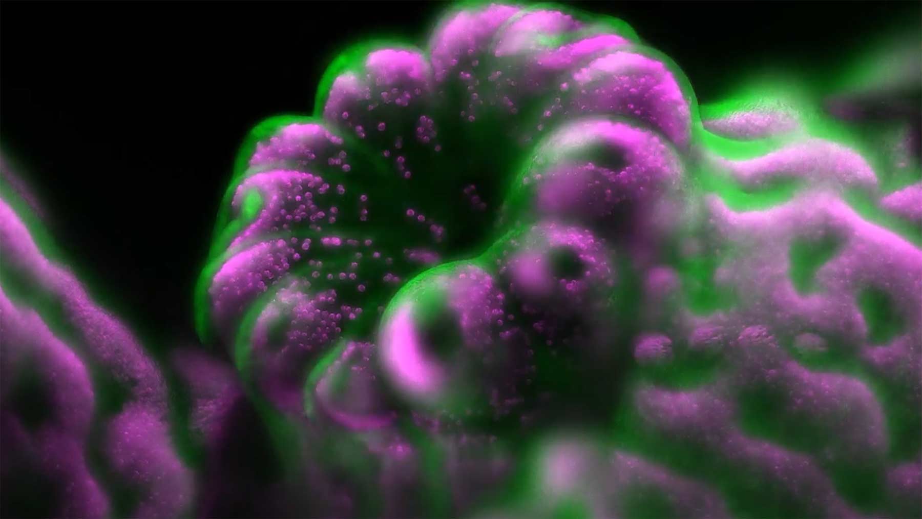
2016 hatte ich mal in Video aus der damaligen Ausgabe der „Nikon Small World in Motion Competition“ gepostet, dieses Mal bin ich etwas näher am Bekanntgabedatum dran und habe zudem gleich die Top-5 der Gewinner-Videos für euch im Angebot. Ob kleinpolypige Steinkorallen-Polypen in Nahaufnahme oder Makro-Aufnahmen eines Mäuse-Embryos – hier wird die Welt der allerkleinsten Teile unter das Mikroskop gelegt und Videokunst daraus gemacht.
„Nikon’s Small World in Motion encompasses any movie or digital time-lapse photography taken through the microscope. Entries are judged by an independent panel of experts who are recognized authorities in the area of photomicrography and photography. These entries are judged on the basis of originality, informational content, technical proficiency and visual impact.“
Platz 5
„Developing mouse embryo, showing the progression of neural tube folding and closure – Dr. Kate McDole & Dr. Philipp Keller“
Platz 4
„Two freshwater tardigrades feeding on another tardigrade – Dr. Hunter N. Hines“
Platz 3
„Stylonychia (microorganism) creating a water vortex using its cilia – Tommy Gunn & Jesse Gunn“
Platz 2
„Vampyrophrya (parasite) tomites swimming rapidly around within the body of the dead copepod host“
Platz 1
„Emerging Acropora muricata (staghorn coral) polyp (coral tissue in green; algae in magenta) – Dr. Philippe P. Laissue“
Viele „Honorable Mentions“ sowie weitere Details zum Wettbewerb, der Jury und den ausgezeichneten Videos findet ihr auf der entsprechenden Projekseite zu den Awards. Alle Videos seit der Initiierung der „Nikon Small World in Motion Competition“ im Jahr 2011 gibt es dort auch zu bestaunen. Schon beachtlich, was sich alles so abspielt, aber von unseren Augen normal gar nicht wahrzunehmen ist. Die Welt ist voller kleiner Wunder. Sehr kleiner Wunder…


Noch keine Kommentare